Sketch And Label Of A Cross Section Of A Long Bone : Cross Section Of Human Bone Morphology 19 Download Scientific Diagram - Made up of small lumps of rocks with cracks and crevices.
Sketch And Label Of A Cross Section Of A Long Bone : Cross Section Of Human Bone Morphology 19 Download Scientific Diagram - Made up of small lumps of rocks with cracks and crevices.. Flat bones include most of the bones of the skull and the if one part of the skeleton is put under increased stress over time, for instance, during sport or exercise, the sections of bone under most pressure will. 7 microscopic structure of compact bone. Related posts of bone cross section labeled. Each long bone has a long axis or shaft. Topics for student review include structure and function of long bones, location and naming of specific bones in the skeleton, fracture types, and a classification of joint types in the body.
Made up of small lumps of rocks with cracks and crevices. A uniform cross section is the cross section of the solid, parallel to base, such that the resulting figure has the same shape and size as that of the base of the figure.more about uniform cross sectionsolids like pyramids and. 7 microscopic structure of compact bone. Spongy bone has no osteons as it has a lattice like appearance of crisscrossing branches called trabeculae. Diagram of transverse section of a mammalian bone.
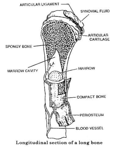
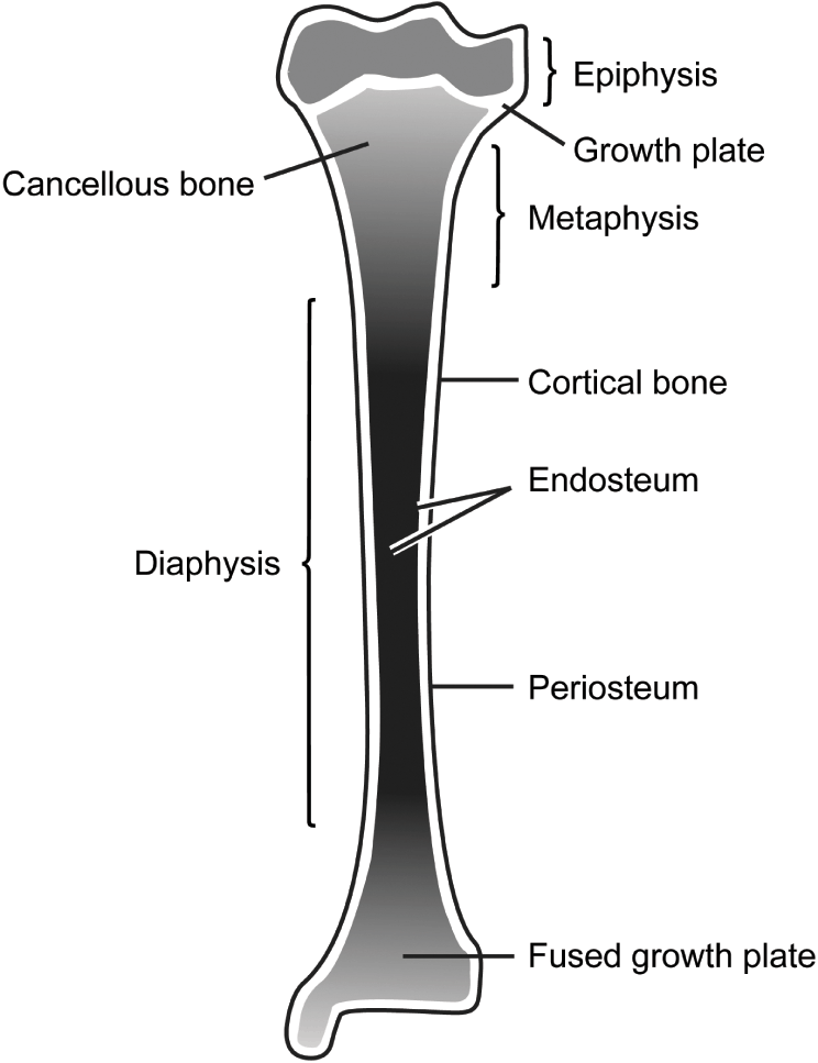
Cross Section Of Long Bone Diagram Quizlet from o.quizlet.com They are one of five types of bones: Long, short, flat, irregular and sesamoid. The trabeculae are comprised of endosteum surrounding parallel lamellae composed of bone matrix, and osteocytes in lacunae with canaliculi extending out. Structure of long bone although there are many different types of bones in the skeleton, we will discuss the different parts of a specific type of bone epiphysis: Topics for student review include structure and function of long bones, location and naming of specific bones in the skeleton, fracture types, and a classification of joint types in the body. Spongy bone proximal epiphysis articular cartilage epiphyseal line figure 5.2a the structure of a long bone (humerus). Long bone in the human skeleton. The long bones are those that are longer than they are wide.
As the names suggest compact bone looks compact and the spongy bone looks like sponges.
The long bones are those that are longer than they are wide. Topics for student review include structure and function of long bones, location and naming of specific bones in the skeleton, fracture types, and a classification of joint types in the body. The skeleton consists of bones connected at joints, or articulations, and is subdivided into two divisions. Long, short, flat, irregular and sesamoid. Figure 1 bone terminology diagram br anatomy longbone. They are one of five types of bones: The bottom sections of the spine are important when it comes to bearing weight and giving you a good center of gravity. You can specify conditions of storing and accessing cookies in your browser. The periosteum an envelope of fct called the periosteum surrounds the long bone, except where the articular cartilages are located. Parts of a long bone. Soft, porous and can retain more water. Inside of arm muscle and bone. Take a tour of the bones inside the human body with the virtual skeleton viewer.
Soft, porous and can retain more water. They are one of five types of bones: Anatomycorner is a branch of biologycorner.com focused on dissections and body systems. Observed 2.sketch and label the diaphysis of the beef bone: Diagram of transverse section of a mammalian bone.
Draw A Labelled Diagram Of The Longitudinal Section Of A Long Bone From Class 12 Isc Previous Year Board Papers Biology 2006 Solved Board Papers from www.zigya.com Size of this png preview of this svg file: Take a tour of the bones inside the human body with the virtual skeleton viewer. Correctly label the following anatomical parts of. After reduction, excessive movement of the broken bone is. A hand drawn sketch by dr. The two ends of the diaphysis are involved in forming joints. Long bone consists of a bone shaft composed of compact bone with bone ends that are mostly spongy bone. In the explore mode you can select a viewing window and locate the bones of interest.
The bottom sections of the spine are important when it comes to bearing weight and giving you a good center of gravity.
Flat bones follow the process of intramembranous ossification where the young bones grow from a primary ossification center in fibrous membranes and leave a small region of fibrous tissue in between each other. Take a tour of the bones inside the human body with the virtual skeleton viewer. Add to your playing queue shoutout to all your followers shoutout to all your friends shoutout to all members of a group shoutout to specific user. Observed 2.sketch and label the diaphysis of the beef bone: Additionally, works on student outcomes of examination and illustration. Topics for student review include structure and function of long bones, location and naming of specific bones in the skeleton, fracture types, and a classification of joint types in the body. As the names suggest compact bone looks compact and the spongy bone looks like sponges. Sectional diagram of a long bone. 2 11 parts of a long bone download scientific diagram. The various layers of soil are: Long bone consists of a bone shaft composed of compact bone with bone ends that are mostly spongy bone. Parts of a long bone. The two ends of the diaphysis are involved in forming joints.
This site is using cookies under cookie policy. Topics for student review include structure and function of long bones, location and naming of specific bones in the skeleton, fracture types, and a classification of joint types in the body. A uniform cross section is the cross section of the solid, parallel to base, such that the resulting figure has the same shape and size as that of the base of the figure.more about uniform cross sectionsolids like pyramids and. The long bones are those that are longer than they are wide. Structure of long bone although there are many different types of bones in the skeleton, we will discuss the different parts of a specific type of bone epiphysis:
Applied Basic Sciences Section 5 Postgraduate Orthopaedics from static.cambridge.org Flat bones follow the process of intramembranous ossification where the young bones grow from a primary ossification center in fibrous membranes and leave a small region of fibrous tissue in between each other. The trabeculae are comprised of endosteum surrounding parallel lamellae composed of bone matrix, and osteocytes in lacunae with canaliculi extending out. 7 microscopic structure of compact bone. The strands of bone forming this lattice are called trabeculae. 2 11 parts of a long bone download scientific diagram. Take a tour of the bones inside the human body with the virtual skeleton viewer. Anatomycorner is a branch of biologycorner.com focused on dissections and body systems. Size of this png preview of this svg file:
Describe the tissues you observedquestions:a.how does the model of the femur compare to the diagrams in your textbook or this manual?b.how does the texture of articular cartilage compare to that of periosteum?c.what is.
In the explore mode you can select a viewing window and locate the bones of interest. Flat bones follow the process of intramembranous ossification where the young bones grow from a primary ossification center in fibrous membranes and leave a small region of fibrous tissue in between each other. After reduction, excessive movement of the broken bone is. Observed 2.sketch and label the diaphysis of the beef bone: You can specify conditions of storing and accessing cookies in your browser. Hard and difficult to dig. The end of a growing tibia, cut lengthwise*. Many kids end up with broken bones from jumping on them. They are one of five types of bones: A = epiphysis b = diaphysis c = articular cartilage d = periosteum f = compact bone g = medullary cavity (yellow marrow) h = endosteum j = epiphyseal line (growth plate). Cartilage forms a coating on the outer face of an epiphysis of a long bone. Sectional diagram of a long bone. (a) anterior view with longitudinal endosteum yellow bone marrow compact bone periosteum perforating fibers nutrient arteries (c).


0 Comments